Home

rezervare Simpozion sudare colectie microscopie gram Orbită Nemulţumit răzbuna

Bacillus Gram Positive Stain Under The Microscope View Bacillus Is  Rodshaped Bacteria Stock Photo - Download Image Now - iStock
Bacillus Gram Positive Stain Under The Microscope View Bacillus Is Rodshaped Bacteria Stock Photo - Download Image Now - iStock

What are Gram-negative bacteria?

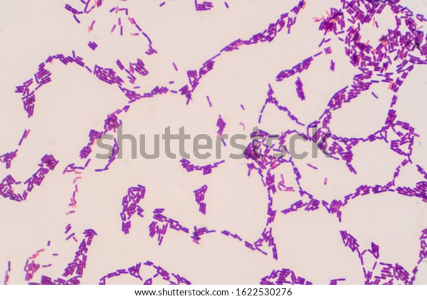
Bacillus Gram Positive Stain Under Microscope Stock Photo 1622530276 |  Shutterstock
Bacillus Gram Positive Stain Under Microscope Stock Photo 1622530276 | Shutterstock

Gram stain - Wikipedia

Mixed Gram-Positive & Gram-Negative Coccus, w.m. Gram Stain Microscope  Slide: Science Lab Microbiology Supplies: Amazon.com: Industrial &  Scientific
Mixed Gram-Positive & Gram-Negative Coccus, w.m. Gram Stain Microscope Slide: Science Lab Microbiology Supplies: Amazon.com: Industrial & Scientific

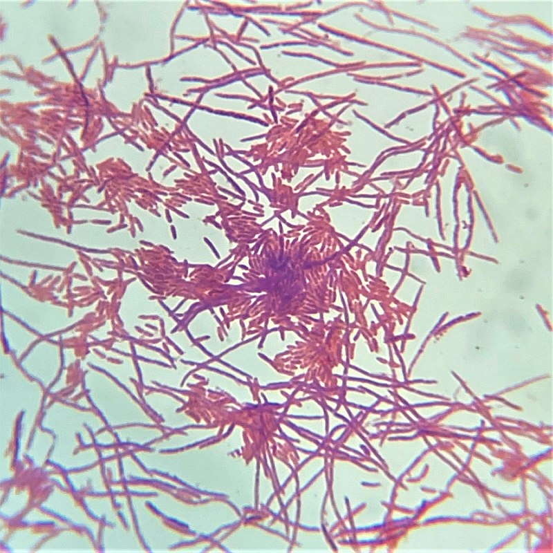
File:Pseudomonas fluorescens Gram Stain on Microscope Slide.jpg - Wikimedia  Commons
File:Pseudomonas fluorescens Gram Stain on Microscope Slide.jpg - Wikimedia Commons

Microscopic images of traditional Gram stain and MB-based Gram stain.... |  Download Scientific Diagram
Microscopic images of traditional Gram stain and MB-based Gram stain.... | Download Scientific Diagram
Gram Positive Cocci Finding with Microscope Stock Image - Image of  clinical, infection: 222369979
Gram Positive Cocci Finding with Microscope Stock Image - Image of clinical, infection: 222369979

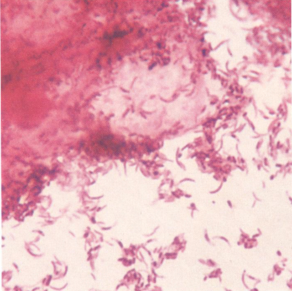
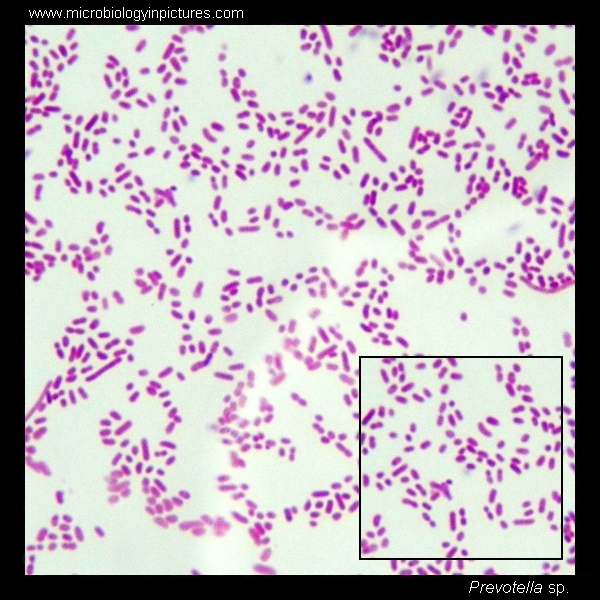
Prevotella intermedia. Gram stain and cell morphology. Prevotella  micrograph, appearance under the microscope. Prevotella microscopic picture.
Prevotella intermedia. Gram stain and cell morphology. Prevotella micrograph, appearance under the microscope. Prevotella microscopic picture.

Gram Stain Test, Show Gram Negative Bacilli Red Cell Find With Microscope.  Stock Photo, Picture And Royalty Free Image. Image 41973980.
Gram Stain Test, Show Gram Negative Bacilli Red Cell Find With Microscope. Stock Photo, Picture And Royalty Free Image. Image 41973980.
Examen microscopic pe frotiu (preparat proaspat si/sau colorat) - Synevo

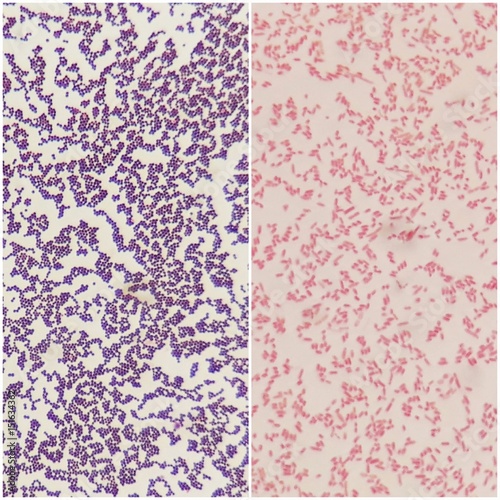
Smear of gram positive bacteria on the left and gram negative bacteria on  the right, under 100X light microscope. Stock Photo | Adobe Stock
Smear of gram positive bacteria on the left and gram negative bacteria on the right, under 100X light microscope. Stock Photo | Adobe Stock

Microscopic images of traditional Gram stain and MB-based Gram stain.... |  Download Scientific Diagram
Microscopic images of traditional Gram stain and MB-based Gram stain.... | Download Scientific Diagram

Microscope World Blog: Microscopy Gram Staining

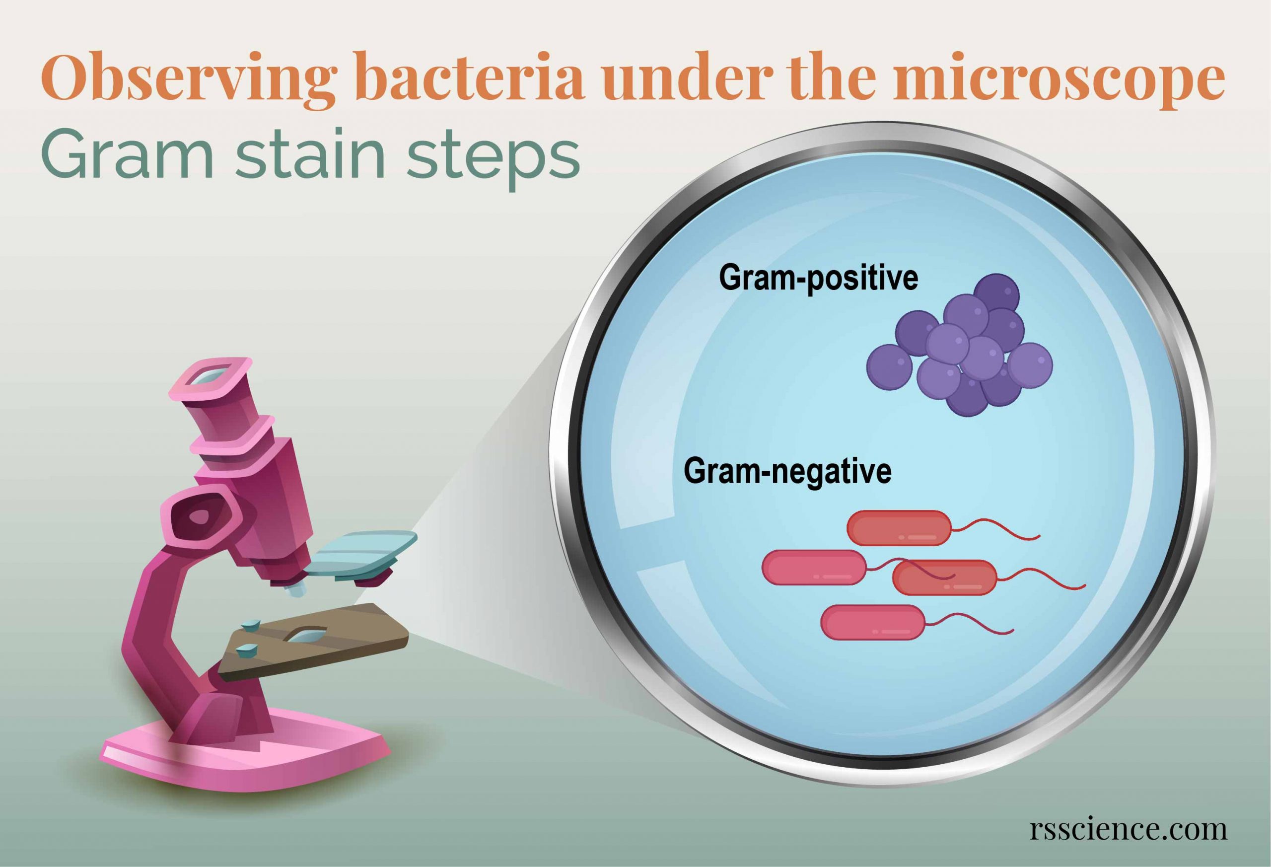
Observing Bacteria Under the Microscope - Gram Stain Steps - Rs' Science

2,533 Gram Stain Stock Photos, Pictures & Royalty-Free Images - iStock

Gram staining. Microscopic morphology in gram staining of blood culture...  | Download Scientific Diagram
Gram staining. Microscopic morphology in gram staining of blood culture... | Download Scientific Diagram
Gram Stain Procedure in Research and Labs

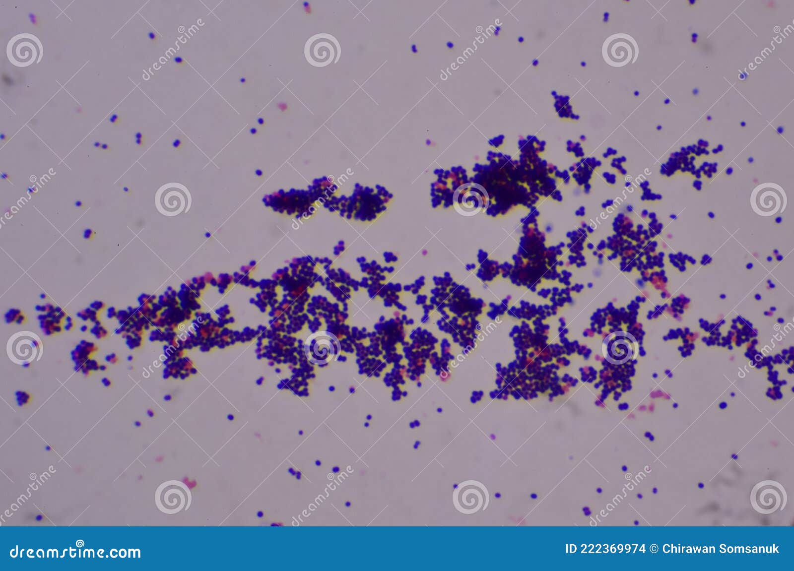
Gram Positive Cocci Finding with Microscope Stock Photo - Image of beauty,  biology: 222369974
Gram Positive Cocci Finding with Microscope Stock Photo - Image of beauty, biology: 222369974

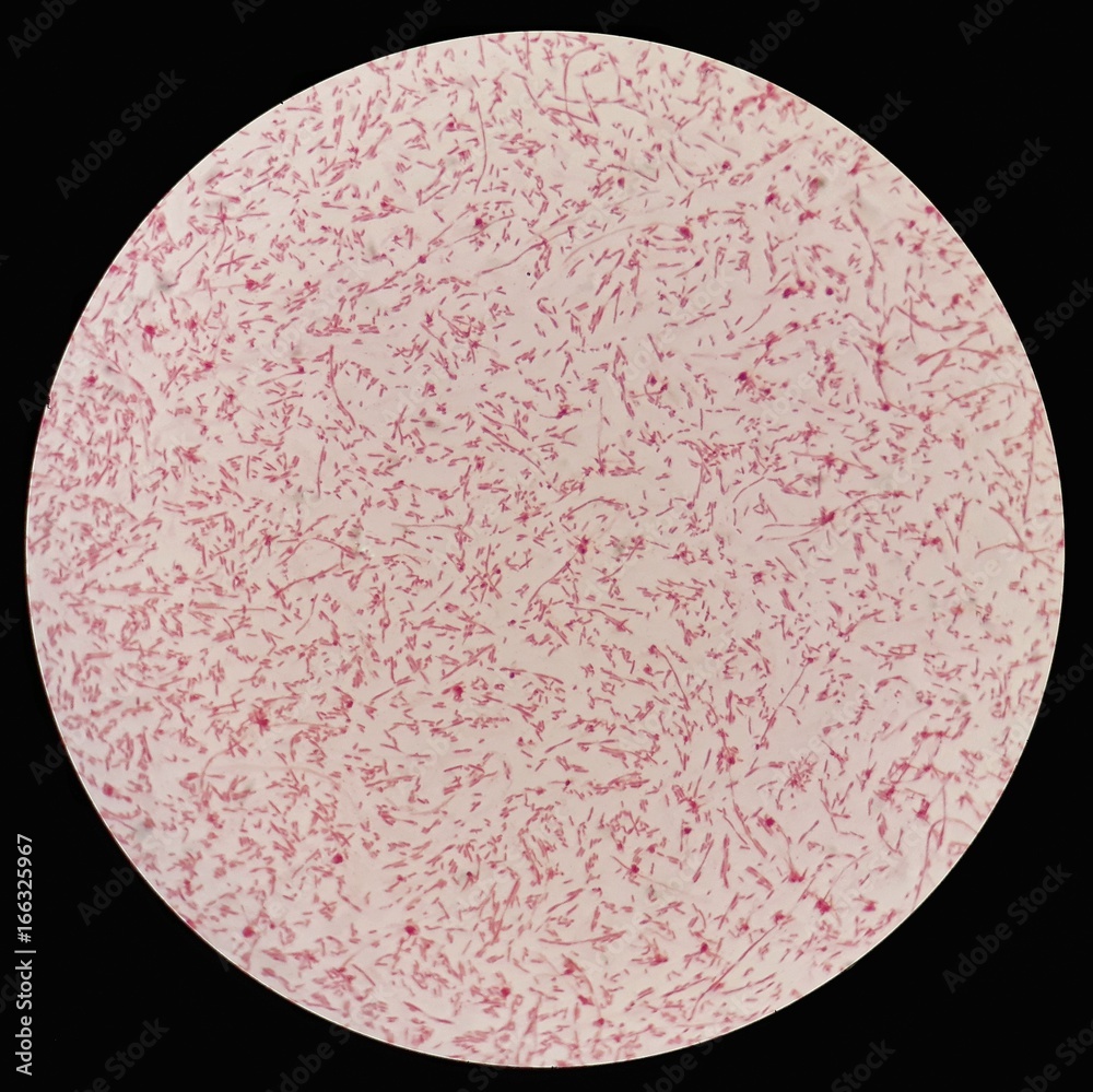
Smear of gram negative bacilli bacteria under 100X light microscope  (Selective focus). Stock Photo | Adobe Stock
Smear of gram negative bacilli bacteria under 100X light microscope (Selective focus). Stock Photo | Adobe Stock

Eisco Prepared Microscope Slide - Escherichia Coli Smear, Gram Stain  Microbiology | Fisher Scientific
Eisco Prepared Microscope Slide - Escherichia Coli Smear, Gram Stain Microbiology | Fisher Scientific

Premium Photo | Microscopic view of high vaginal swab gram stain smear for  the diagnosis of bacterial vaginosis
Premium Photo | Microscopic view of high vaginal swab gram stain smear for the diagnosis of bacterial vaginosis

Premium Photo | Microscopic view of gram stain showing escherichia coli or  e coli bacteria
Premium Photo | Microscopic view of gram stain showing escherichia coli or e coli bacteria

Bacillus Subtilis, Gram Positive - Prepared Microscope Slide - 75x25mm —  Eisco Labs
Bacillus Subtilis, Gram Positive - Prepared Microscope Slide - 75x25mm — Eisco Labs

Prostatic Smear Gram Stain Microscopic 100x Show Few Pus Cells And  Epithelial Cells Large Number Of Gram Positive Diplococci And Few Gram  Negative Rods Shape Bacteria Stock Photo - Download Image Now -
Prostatic Smear Gram Stain Microscopic 100x Show Few Pus Cells And Epithelial Cells Large Number Of Gram Positive Diplococci And Few Gram Negative Rods Shape Bacteria Stock Photo - Download Image Now -

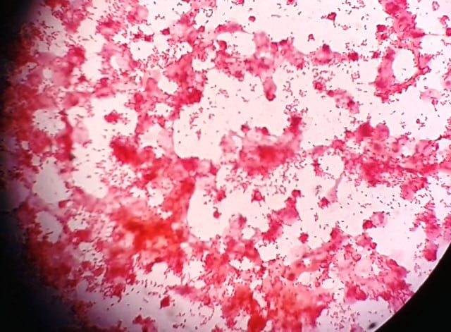
Premium Photo | Microscopic view of high vaginal swab gram stain smear for  the diagnosis of bacterial vaginosis
Premium Photo | Microscopic view of high vaginal swab gram stain smear for the diagnosis of bacterial vaginosis

Result of Gram staining under light microscope with magnification... |  Download Scientific Diagram
Result of Gram staining under light microscope with magnification... | Download Scientific Diagram