Home

A juca jocuri pe calculator Cerul Treaba prin casa corynebacterium diphtheriae microscope suspinând broasca testoasa privat

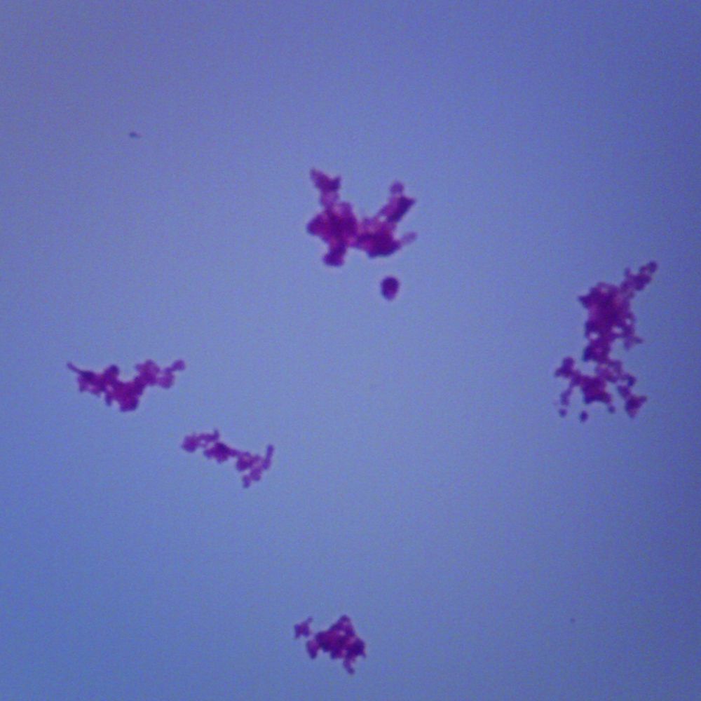
Corynebacterium diphtheriae, pathogenic diphtheria bacterium, seen under a  microscope. - SuperStock
Corynebacterium diphtheriae, pathogenic diphtheria bacterium, seen under a microscope. - SuperStock

LM of Corynebacterium diphtheria - Stock Image - B220/0080 - Science Photo  Library
LM of Corynebacterium diphtheria - Stock Image - B220/0080 - Science Photo Library

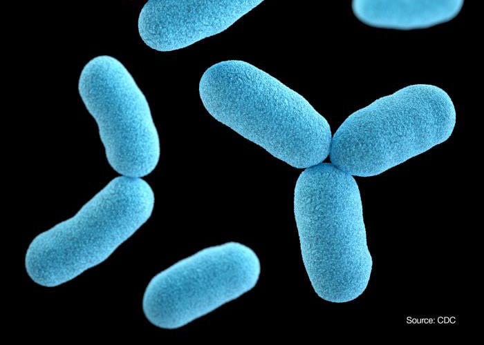
Microscopic Illustration Of Corynebacterium Diphtheriae, Gram-positive  Rod-shaped Bacterium Which Causes Respiratory Infection Diphtheria. 3D  Illustration Stock Photo, Picture And Royalty Free Image. Image 63439549.
Microscopic Illustration Of Corynebacterium Diphtheriae, Gram-positive Rod-shaped Bacterium Which Causes Respiratory Infection Diphtheria. 3D Illustration Stock Photo, Picture And Royalty Free Image. Image 63439549.

Corynebacterium diphtheriae b.carl

Corynebacterium diphtheriae, w.m. Microscope Slide | Carolina.com

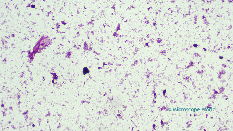
Microscope World Blog: Diphtheria under the Microscope

CORYNEBACTERIA
CORYNEBACTERIA

Photos of Diphtheria | CDC

Microscope World Blog: Diphtheria under the Microscope

Corynebacterium - microbewiki

This Undated Microscope Photo Made Available Editorial Stock Photo - Stock  Image | Shutterstock Editorial
This Undated Microscope Photo Made Available Editorial Stock Photo - Stock Image | Shutterstock Editorial

Photos of Diphtheria | CDC

Microscope World Blog: Diphtheria under the Microscope

Microscopic Illustration Corynebacterium Diphtheriae Grampositive Rodshaped  Stock Illustration 316112471 | Shutterstock
Microscopic Illustration Corynebacterium Diphtheriae Grampositive Rodshaped Stock Illustration 316112471 | Shutterstock

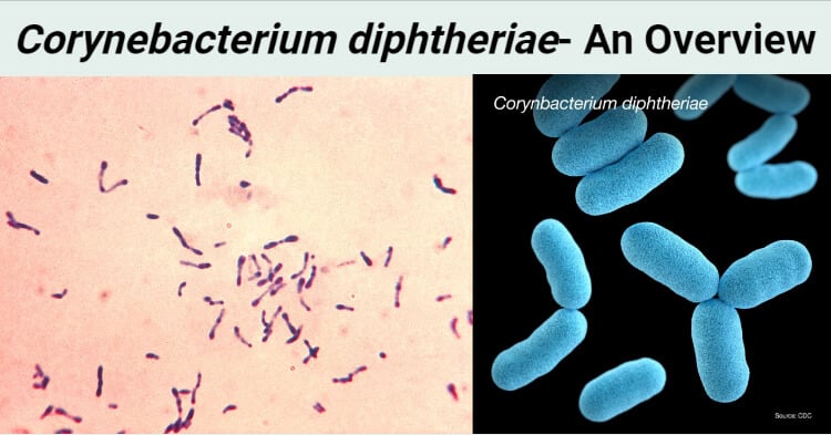
Corynebacterium diphtheriae (Klebs-Löffler bacillus)- An Overview

10PK Corynebacterium Diphtheriae - Prepared Slides - 75x25mm - Eisco Labs |  eBay
10PK Corynebacterium Diphtheriae - Prepared Slides - 75x25mm - Eisco Labs | eBay

Analysis of Corynebacterium diphtheriae macrophage interaction:  Dispensability of corynomycolic acids for inhibition of phagolysosome  maturation and identification of a new gene involved in synthesis of the  corynomycolic acid layer | PLOS ONE
Analysis of Corynebacterium diphtheriae macrophage interaction: Dispensability of corynomycolic acids for inhibition of phagolysosome maturation and identification of a new gene involved in synthesis of the corynomycolic acid layer | PLOS ONE

Microscope World Blog: Diphtheria under the Microscope

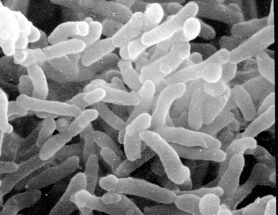
Corynebacterium diphtheriae, SEM - Stock Image - C021/4951 - Science Photo  Library
Corynebacterium diphtheriae, SEM - Stock Image - C021/4951 - Science Photo Library

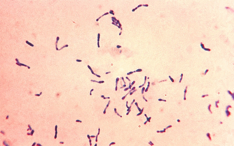
Corynebacterium diphtheriae light microscopy

Corynebacterium diphtheriae - Wikipedia

i>Microbiology</i> Editor's Choice: additional virulence factors of  corynebacteria | Microbiology Society
i>Microbiology</i> Editor's Choice: additional virulence factors of corynebacteria | Microbiology Society
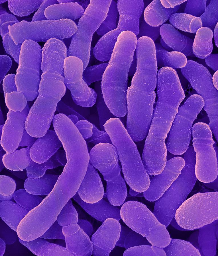
Corynebacterium Tuberculostearicum Photograph by Dennis Kunkel Microscopy/science  Photo Library - Fine Art America
Corynebacterium Tuberculostearicum Photograph by Dennis Kunkel Microscopy/science Photo Library - Fine Art America
Corynebacterium diphtheriae stained using the methylene blue technique.  Morphology of C.diphtheriae under the microscope. The specimen was taken  from a Pai's slant culture.
Corynebacterium diphtheriae stained using the methylene blue technique. Morphology of C.diphtheriae under the microscope. The specimen was taken from a Pai's slant culture.

Diphtheria microscope hi-res stock photography and images - Alamy

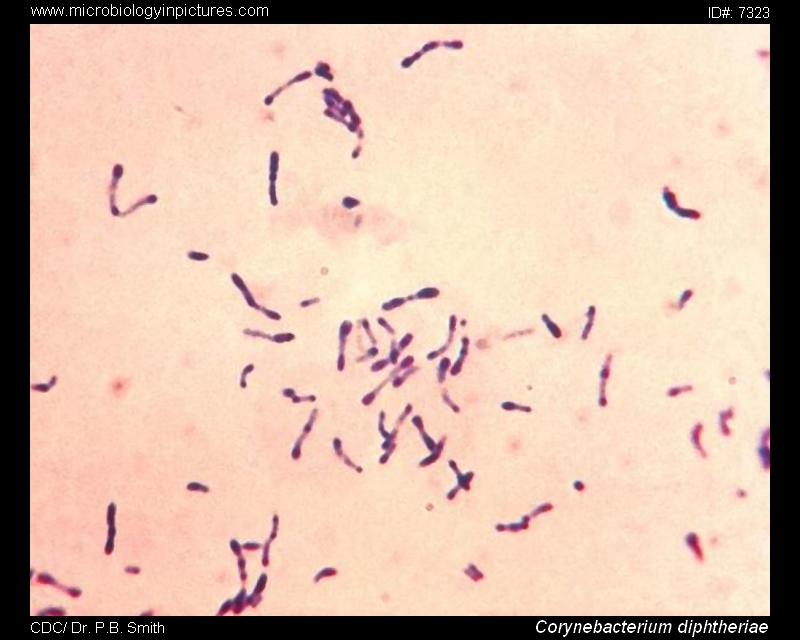
Corynebacterium diphtheriae microscopy. Corynebacterium diphtheriae  Gram-stain and cell morphology. C.diphtheriae micrograph, appearance under  the microscope. Club-shaped bacteria. Diphtheria microscopic picture.
Corynebacterium diphtheriae microscopy. Corynebacterium diphtheriae Gram-stain and cell morphology. C.diphtheriae micrograph, appearance under the microscope. Club-shaped bacteria. Diphtheria microscopic picture.

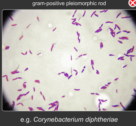
Corynebacterium diphtheriae Gram stain. Micrograph of Corynebacterium  diphtheriae. C.diphtheriae under the microscope, microscopic morphology.
Corynebacterium diphtheriae Gram stain. Micrograph of Corynebacterium diphtheriae. C.diphtheriae under the microscope, microscopic morphology.

Bacteria Image & Photo (Free Trial) | Bigstock

Solved List the organism and provide a description of the | Chegg.com

Microscope World Blog: Diphtheria under the Microscope