Home

Balena Baleen Spuneți deoparte presiune ganglion corti parc Primar bufet

ファイル:Organ of corti.svg - Wikipedia

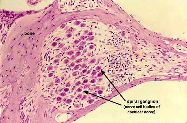
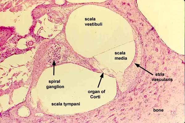
Histology at SIU
Histology at SIU

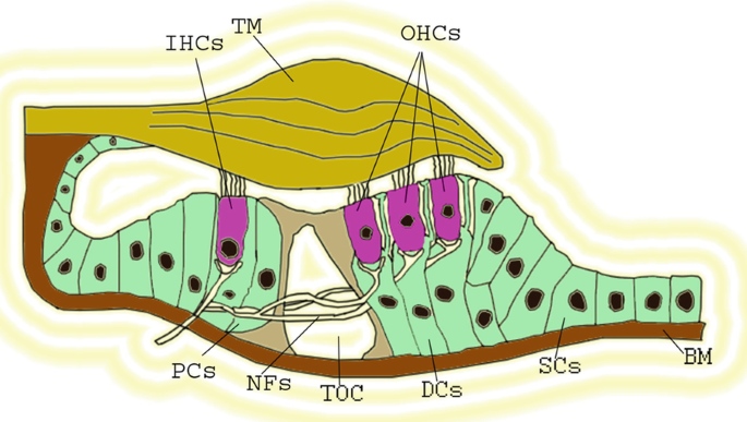
Function of the Organ of Corti

Type II spiral ganglion afferent neurons drive medial olivocochlear reflex  suppression of the cochlear amplifier | Nature Communications
Type II spiral ganglion afferent neurons drive medial olivocochlear reflex suppression of the cochlear amplifier | Nature Communications

Prickle1 is expressed in the spiral ganglion but not the organ of Corti...  | Download Scientific Diagram
Prickle1 is expressed in the spiral ganglion but not the organ of Corti... | Download Scientific Diagram

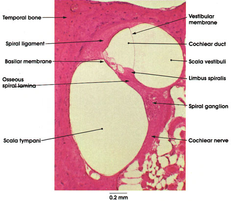
Anatomy Atlases: Atlas of Microscopic Anatomy: Section 1 - Cells

Schwann cells revert to non‐myelinating phenotypes in the deafened rat  cochlea - Hurley - 2007 - European Journal of Neuroscience - Wiley Online  Library
Schwann cells revert to non‐myelinating phenotypes in the deafened rat cochlea - Hurley - 2007 - European Journal of Neuroscience - Wiley Online Library

Spike Encoding of Neurotransmitter Release Timing by Spiral Ganglion  Neurons of the Cochlea | Journal of Neuroscience
Spike Encoding of Neurotransmitter Release Timing by Spiral Ganglion Neurons of the Cochlea | Journal of Neuroscience

Approaches to Regenerate Hair Cell and Spiral Ganglion Neuron in the Inner  Ear | SpringerLink
Approaches to Regenerate Hair Cell and Spiral Ganglion Neuron in the Inner Ear | SpringerLink

Auditory neurons form the spiral ganglion in the cochlea and connect... |  Download Scientific Diagram
Auditory neurons form the spiral ganglion in the cochlea and connect... | Download Scientific Diagram

The Auditory Pathway - Structures of the Ear - Auditory Transduction -  TeachMeAnatomy
The Auditory Pathway - Structures of the Ear - Auditory Transduction - TeachMeAnatomy

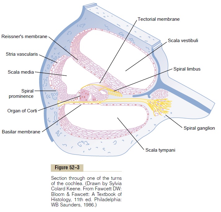
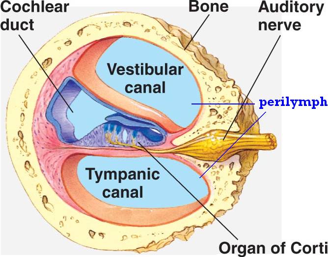
Cross-section through the mammalian cochlea showing the three cochlear... |  Download Scientific Diagram
Cross-section through the mammalian cochlea showing the three cochlear... | Download Scientific Diagram
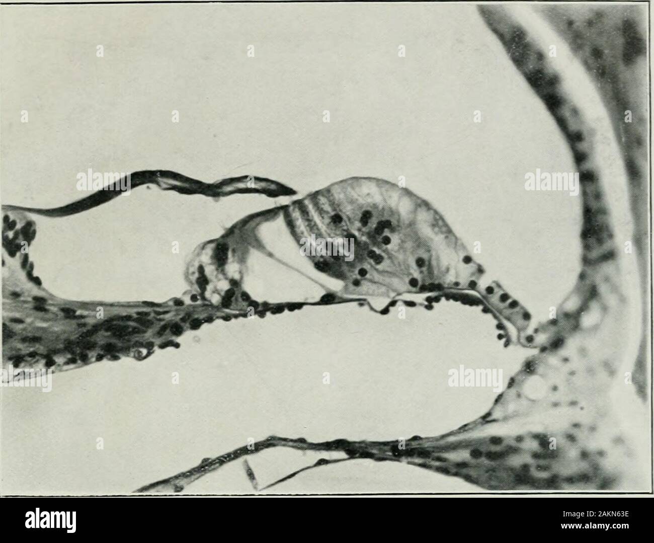
The Journal of laryngology and otology . Fig. 6.—Organ of Corti of white  iuoii.se. i lon. Showing the delicate rodsof Corti and the short outer  hair-cells. The ganglion spirale is seen tothe
The Journal of laryngology and otology . Fig. 6.—Organ of Corti of white iuoii.se. i lon. Showing the delicate rodsof Corti and the short outer hair-cells. The ganglion spirale is seen tothe
Organ of Corti in the ear stock vector. Illustration of tympani - 60932666

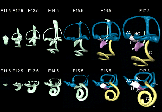
Inner ear development: building a spiral ganglion and an organ of Corti out  of unspecified ectoderm | SpringerLink
Inner ear development: building a spiral ganglion and an organ of Corti out of unspecified ectoderm | SpringerLink
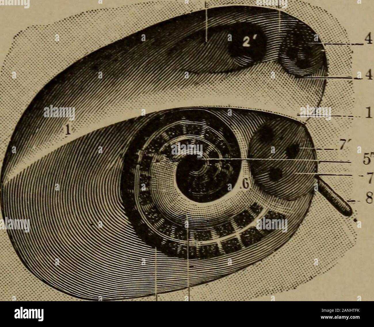
Spiral ganglion hi-res stock photography and images - Alamy

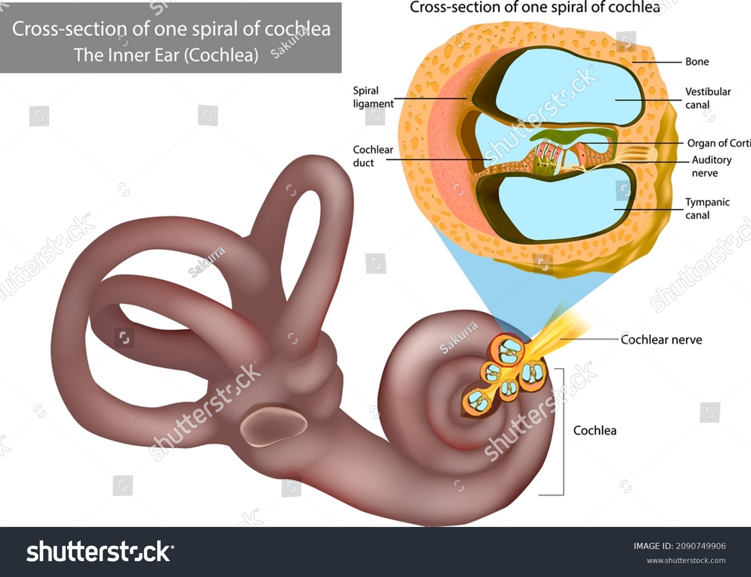
22 Spiral Ganglion Images, Stock Photos & Vectors | Shutterstock

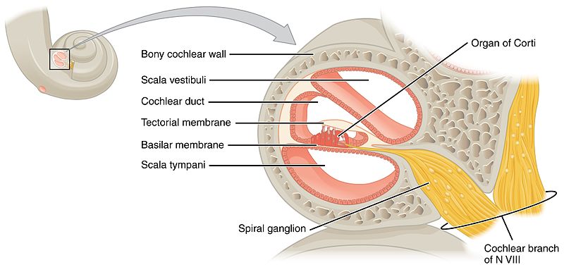
Organ of Corti
Organ of Corti

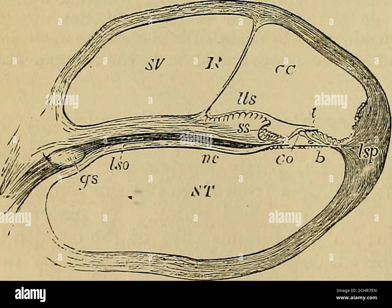
Quain's elements of anatomy . piralis. scala tympani (ST), but does not  properly speaking enter into the lowerboundary of the scala vestibuh, for a  second, much more delicate mem- Fig. 398.
Quain's elements of anatomy . piralis. scala tympani (ST), but does not properly speaking enter into the lowerboundary of the scala vestibuh, for a second, much more delicate mem- Fig. 398.

The organization of SGN innervation in the mature cochlea. a, In a... |  Download Scientific Diagram
The organization of SGN innervation in the mature cochlea. a, In a... | Download Scientific Diagram

Altered expression of genes regulating inflammation and synaptogenesis  during regrowth of afferent neurons to cochlear hair cells | PLOS ONE
Altered expression of genes regulating inflammation and synaptogenesis during regrowth of afferent neurons to cochlear hair cells | PLOS ONE

Capturing PGCs in the human cochlea. (A) Schematic model of PGC... |  Download Scientific Diagram
Capturing PGCs in the human cochlea. (A) Schematic model of PGC... | Download Scientific Diagram

Prickle1 regulates neurite outgrowth of apical spiral ganglion neurons but  not hair cell polarity in the murine cochlea | PLOS ONE
Prickle1 regulates neurite outgrowth of apical spiral ganglion neurons but not hair cell polarity in the murine cochlea | PLOS ONE

Why do hair cells and spiral ganglion neurons in the cochlea die during  aging? - Abstract - Europe PMC
Why do hair cells and spiral ganglion neurons in the cochlea die during aging? - Abstract - Europe PMC

Spiral ganglion - Wikipedia

Histology at SIU, ear