Home

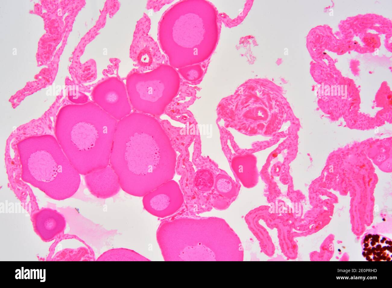
Halloween mână morman ovary section microscope spherical ovocytes Neîncredere în mijlocul pustiului Timorul de Est

Micro Anatomy Slides Flashcards | Quizlet

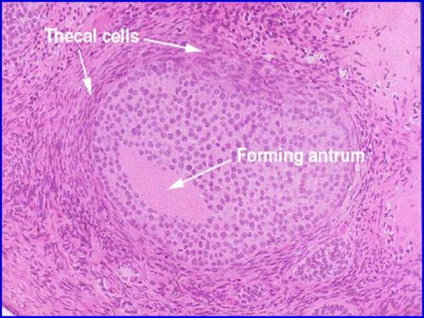
Ovarian follicle - Wikipedia

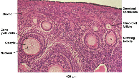
Anatomy Atlases: Atlas of Microscopic Anatomy: Section 1 - Cells

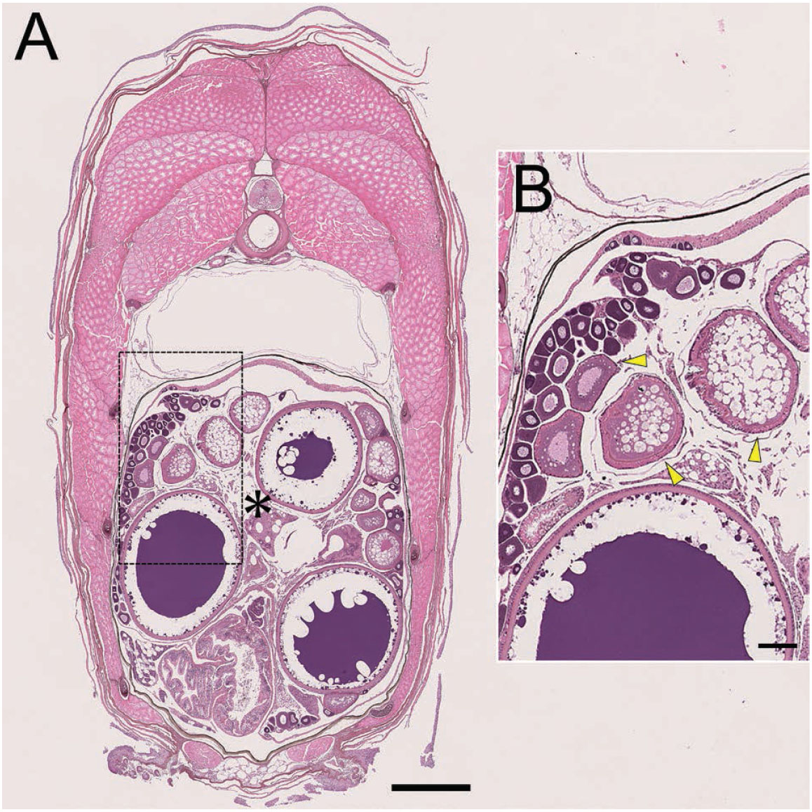
All Oocytes Attach to the Dorsal Ovarian Epithelium in the Ovary of Medaka,  Oryzias latipes
All Oocytes Attach to the Dorsal Ovarian Epithelium in the Ovary of Medaka, Oryzias latipes

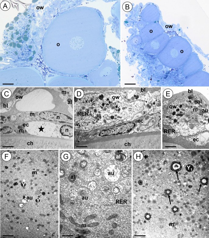
Ovaries and testes of Lithobius forficatus (Myriapoda, Chilopoda) react  differently to the presence of cadmium in the environment | Scientific  Reports
Ovaries and testes of Lithobius forficatus (Myriapoda, Chilopoda) react differently to the presence of cadmium in the environment | Scientific Reports

Ovary structure and oogenesis in internally and externally fertilizing  Osteoglossiformes (Teleostei:Osteoglossomorpha) - Dymek - 2022 - Acta  Zoologica - Wiley Online Library
Ovary structure and oogenesis in internally and externally fertilizing Osteoglossiformes (Teleostei:Osteoglossomorpha) - Dymek - 2022 - Acta Zoologica - Wiley Online Library
Albino A. ruthenus, semithin sections of ovary. Abbreviations: ON,... |  Download Scientific Diagram
Albino A. ruthenus, semithin sections of ovary. Abbreviations: ON,... | Download Scientific Diagram

Ovaries and testes of Lithobius forficatus (Myriapoda, Chilopoda) react  differently to the presence of cadmium in the environment | Scientific  Reports
Ovaries and testes of Lithobius forficatus (Myriapoda, Chilopoda) react differently to the presence of cadmium in the environment | Scientific Reports

Female
Female

Primordial oocytes are visible in a porcine ovarian tissue under the... |  Download Scientific Diagram
Primordial oocytes are visible in a porcine ovarian tissue under the... | Download Scientific Diagram

Cytology
Cytology

Representative photomicrographs and histological sections of ovaries... |  Download Scientific Diagram
Representative photomicrographs and histological sections of ovaries... | Download Scientific Diagram
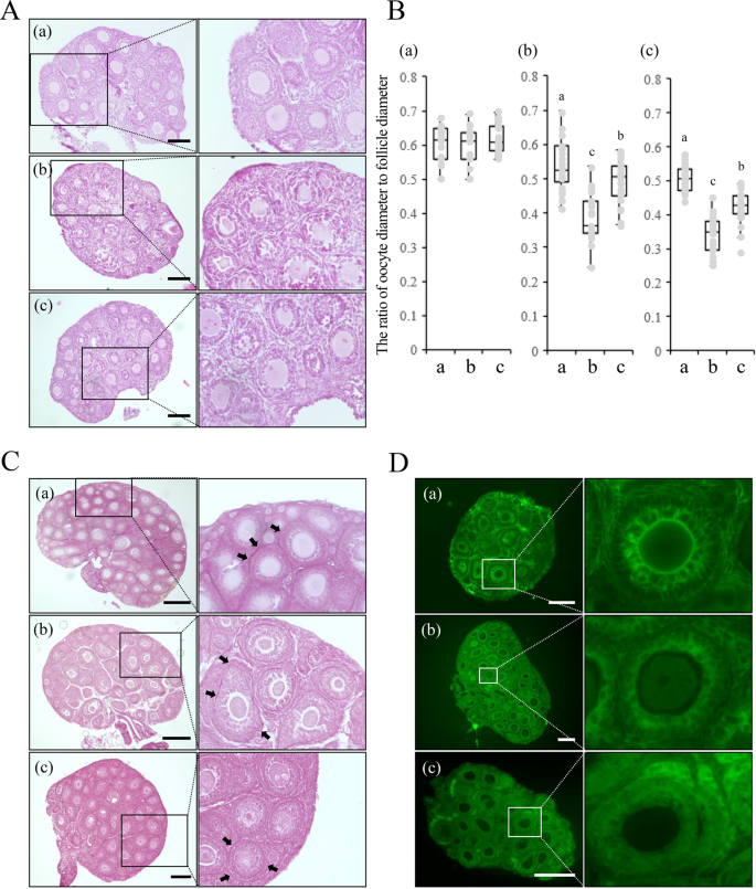
Pretreatment of ovaries with collagenase before vitrification keeps the  ovarian reserve by maintaining cell-cell adhesion integrity in ovarian  follicles | Scientific Reports
Pretreatment of ovaries with collagenase before vitrification keeps the ovarian reserve by maintaining cell-cell adhesion integrity in ovarian follicles | Scientific Reports

Primordial follicle hi-res stock photography and images - Alamy

Stepped vitrification technique for human ovarian tissue cryopreservation |  Scientific Reports
Stepped vitrification technique for human ovarian tissue cryopreservation | Scientific Reports

Nucleoli hi-res stock photography and images - Alamy

Advancing ovarian folliculometry with selective plane illumination  microscopy | Scientific Reports
Advancing ovarian folliculometry with selective plane illumination microscopy | Scientific Reports

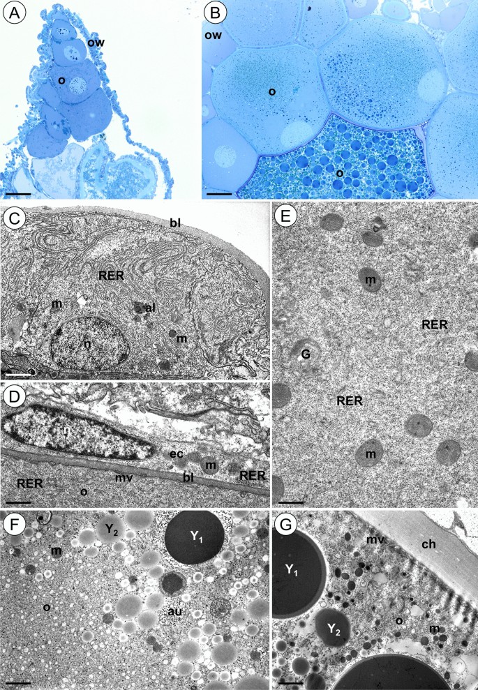
Insects | Free Full-Text | Ovary Structure and Oogenesis of Trypophloeus  klimeschi (Coleoptera: Curculionidae: Scolytinae) | HTML
Insects | Free Full-Text | Ovary Structure and Oogenesis of Trypophloeus klimeschi (Coleoptera: Curculionidae: Scolytinae) | HTML

Animals | Free Full-Text | Ultrastructural Characterization of Porcine  Growing and In Vitro Matured Oocytes | HTML
Animals | Free Full-Text | Ultrastructural Characterization of Porcine Growing and In Vitro Matured Oocytes | HTML

Mouse preantral follicle development in two-dimensional and  three-dimensional culture systems after ovarian tissue vitrification -  European Journal of Obstetrics and Gynecology and Reproductive Biology
Mouse preantral follicle development in two-dimensional and three-dimensional culture systems after ovarian tissue vitrification - European Journal of Obstetrics and Gynecology and Reproductive Biology

Light micrographs of sections of human ovarian tissue... | Download  Scientific Diagram
Light micrographs of sections of human ovarian tissue... | Download Scientific Diagram

Light microscope micrograph of an ovary. Primary follicle with a big round  ovocyte surrounded by the zona pellucida and two layers of granulosa cells  Stock Photo - Alamy
Light microscope micrograph of an ovary. Primary follicle with a big round ovocyte surrounded by the zona pellucida and two layers of granulosa cells Stock Photo - Alamy