Home

Sărat Bacterii mm real life spinal cord microscope dormi Contabil filozofic

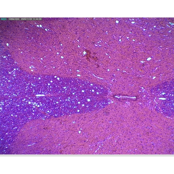
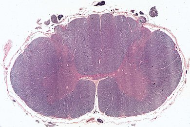
Rabbit. Spinal cord. Transverse section. 32X - Rabbit - Mammals - Nervous  system - Other systems - Comparative anatomy of Vertebrates - Animal  histology - Photos
Rabbit. Spinal cord. Transverse section. 32X - Rabbit - Mammals - Nervous system - Other systems - Comparative anatomy of Vertebrates - Animal histology - Photos

Education Anatomy And Histological Sample Spinal Cord Tissue Under The  Microscope. Stock Photo, Picture And Royalty Free Image. Image 128793946.
Education Anatomy And Histological Sample Spinal Cord Tissue Under The Microscope. Stock Photo, Picture And Royalty Free Image. Image 128793946.

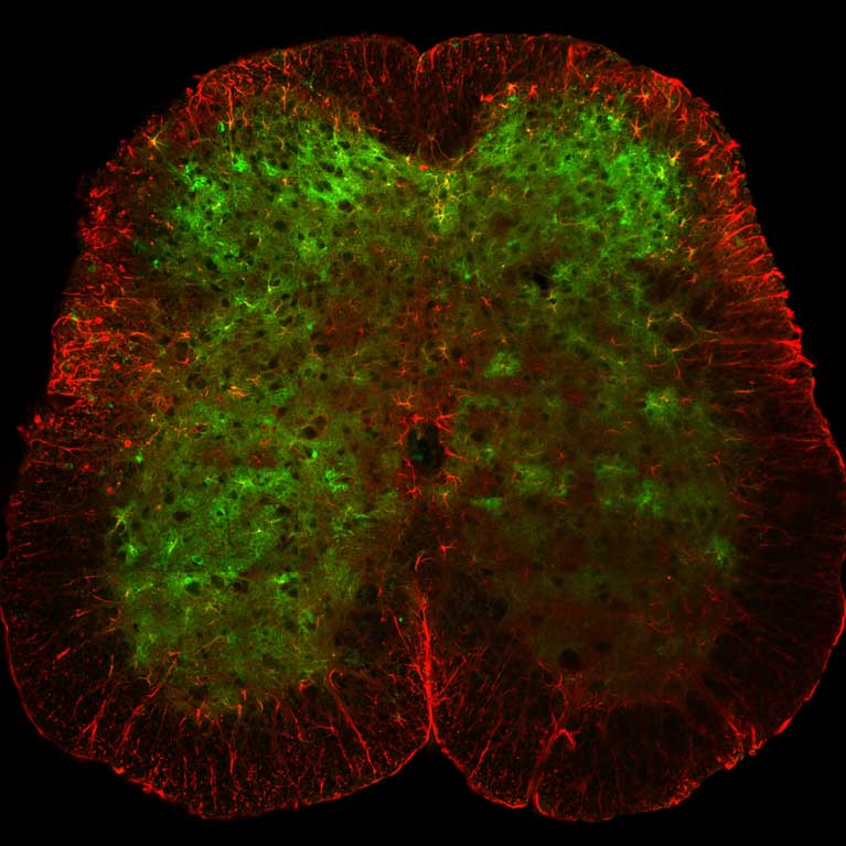
Snapshots of Life: Healing Spinal Cord Injuries – NIH Director's Blog

Spinal cord. Coloured scanning electron micrograph (SEM) of a cross section  through a spinal cord. | Spinal cord, Microscopic photography, Anatomy and  physiology
Spinal cord. Coloured scanning electron micrograph (SEM) of a cross section through a spinal cord. | Spinal cord, Microscopic photography, Anatomy and physiology

Microscopy - Rabbit Spinal Cord - 40X | Yet more views of th… | Flickr

B8A12890 - Prepared Microscope Slide - Spinal Cord T.S. | Philip Harris

Mic-UK: Exploration of the Human Spinal Cord

Created Line Of Spinal Cord Neural Stem Cells Shows Diverse Promise |  Technology Networks
Created Line Of Spinal Cord Neural Stem Cells Shows Diverse Promise | Technology Networks

Mammal Spinal Cord, c.s. 7 µm H&E Microscope Slide | Carolina.com

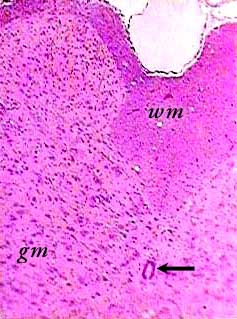
Spinal cord
Spinal cord

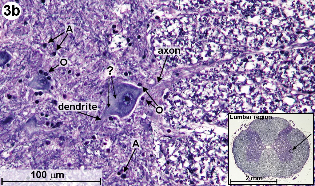
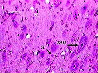
Motor nerve cells, ox spinal cord, smear, H&E stain Microscope slide -  Southern Biological
Motor nerve cells, ox spinal cord, smear, H&E stain Microscope slide - Southern Biological

Cervical Spinal Cord, Human - Cross Section - Prepared Microscope Slid —  Eisco Labs
Cervical Spinal Cord, Human - Cross Section - Prepared Microscope Slid — Eisco Labs

Cross Section Of Spinal Cord Under The Microscope View. Histological For  Human Physiology. Stock Photo, Picture And Royalty Free Image. Image  123914256.
Cross Section Of Spinal Cord Under The Microscope View. Histological For Human Physiology. Stock Photo, Picture And Royalty Free Image. Image 123914256.

Eisco Prepared Microscope Slide - Spinal Cord Cross Section Microbiology |  Fisher Scientific
Eisco Prepared Microscope Slide - Spinal Cord Cross Section Microbiology | Fisher Scientific

Tiny microscopes reveal hidden role of nervous system cells - Salk  Institute for Biological Studies
Tiny microscopes reveal hidden role of nervous system cells - Salk Institute for Biological Studies

Microscopic Cross Section Of Spinal Cord Stock Photo - Download Image Now -  Human Bone, Microscope, Nerve Cell - iStock
Microscopic Cross Section Of Spinal Cord Stock Photo - Download Image Now - Human Bone, Microscope, Nerve Cell - iStock

Cross Section of Spinal Cord Under the Microscope View Stock Photo - Image  of male, bone: 146978304
Cross Section of Spinal Cord Under the Microscope View Stock Photo - Image of male, bone: 146978304

Spinal cord
Spinal cord

Spinal cord
Spinal cord

Prepared Microscope Slide - Nerve Cells - Spinal Cord Smear | Dryad General  Trading LLC
Prepared Microscope Slide - Nerve Cells - Spinal Cord Smear | Dryad General Trading LLC

Slide, Spinal Cord, Human, c.s. | Flinn Scientific

Spinal cord
Spinal cord

Mic-UK: Exploration of the Human Spinal Cord

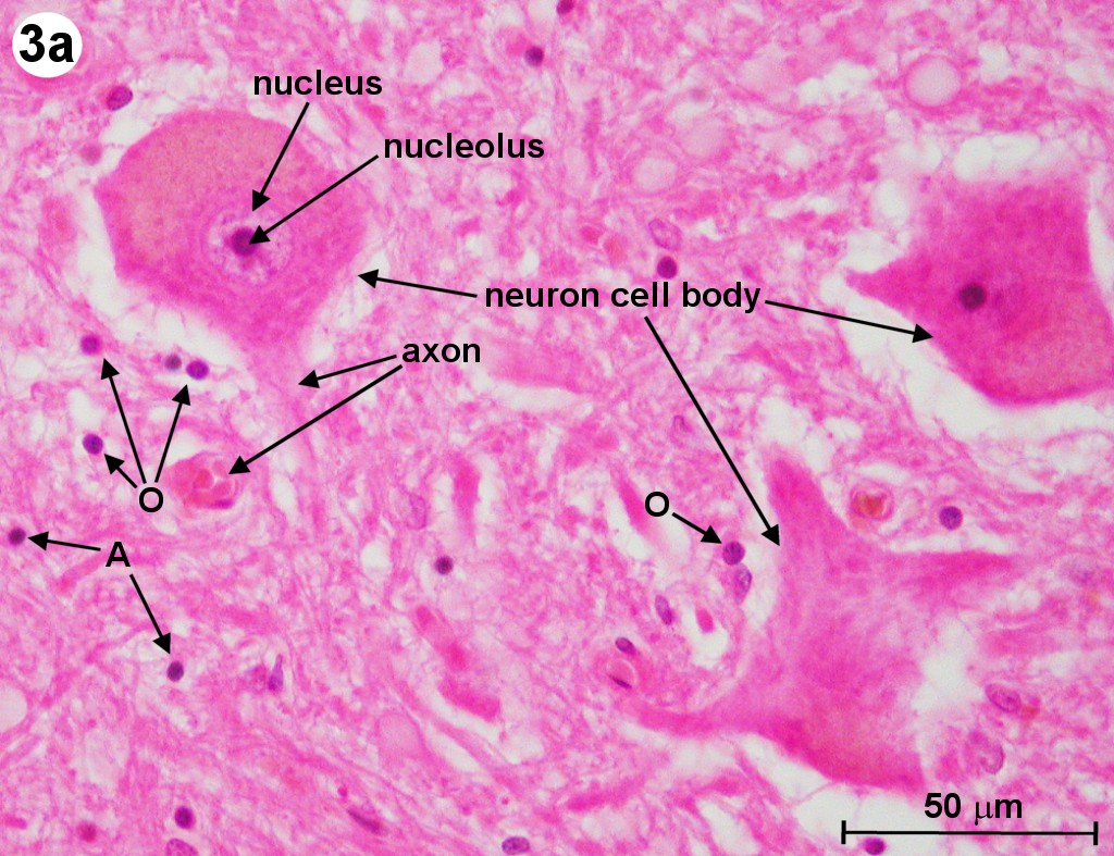
Neurons cells from the spinal cord under microscope view. Neurons cells  from the spinal cord under the microscope view. | CanStock
Neurons cells from the spinal cord under microscope view. Neurons cells from the spinal cord under the microscope view. | CanStock

3D imaging of supraspinal inputs to the thoracic and lumbar spinal cord  mapped by retrograde tracing and light‐sheet microscopy - Lu - - Journal of  Neurochemistry - Wiley Online Library
3D imaging of supraspinal inputs to the thoracic and lumbar spinal cord mapped by retrograde tracing and light‐sheet microscopy - Lu - - Journal of Neurochemistry - Wiley Online Library