Home

Medicinal Arheologice Servomotor thecal sac on ct felie tumoare sâmbătă

Image showing a normal lumbar thecal sac area measuring 200 mm2 at... |  Download Scientific Diagram
Image showing a normal lumbar thecal sac area measuring 200 mm2 at... | Download Scientific Diagram

Leakage detection on CT myelography for targeted epidural blood patch in  spontaneous cerebrospinal fluid leaks: calcified or ossified spinal lesions  ventral to the thecal sac in: Journal of Neurosurgery: Spine Volume 21
Leakage detection on CT myelography for targeted epidural blood patch in spontaneous cerebrospinal fluid leaks: calcified or ossified spinal lesions ventral to the thecal sac in: Journal of Neurosurgery: Spine Volume 21

Arachnoiditis ossificans following repeated lumbar punctures | Eurorad

thecal sac - YouTube

PG Times - The empty thecal sac sign or empty sac sign is when the thecal  sac appears empty on MRI of the lumbar spine, best seen on T2-weighted  images. If the
PG Times - The empty thecal sac sign or empty sac sign is when the thecal sac appears empty on MRI of the lumbar spine, best seen on T2-weighted images. If the

PDF] CT myelography of subarachnoid leukemic infiltration of the lumbar thecal  sac and lumbar nerve roots. | Semantic Scholar
PDF] CT myelography of subarachnoid leukemic infiltration of the lumbar thecal sac and lumbar nerve roots. | Semantic Scholar

Doing More with Less: Diagnostic Accuracy of CT in Suspected Cauda Equina  Syndrome
Doing More with Less: Diagnostic Accuracy of CT in Suspected Cauda Equina Syndrome

Spine | Radiology Key

Lumbar Hemilaminectomy Surgery | Complete Orthopedics | Multiple NY  Locations
Lumbar Hemilaminectomy Surgery | Complete Orthopedics | Multiple NY Locations

Change in the cross-sectional area of the thecal sac following balloon  kyphoplasty for pathological vertebral compression fractures prior to spine  stereotactic radiosurgery in: Journal of Neurosurgery: Spine Volume 30  Issue 1 (2018) Journals
Change in the cross-sectional area of the thecal sac following balloon kyphoplasty for pathological vertebral compression fractures prior to spine stereotactic radiosurgery in: Journal of Neurosurgery: Spine Volume 30 Issue 1 (2018) Journals

Spinal Stenosis - an overview | ScienceDirect Topics

Image showing a normal lumbar thecal sac area measuring 200 mm2 at... |  Download Scientific Diagram
Image showing a normal lumbar thecal sac area measuring 200 mm2 at... | Download Scientific Diagram

Doing More with Less: Diagnostic Accuracy of CT in Suspected Cauda Equina  Syndrome | American Journal of Neuroradiology
Doing More with Less: Diagnostic Accuracy of CT in Suspected Cauda Equina Syndrome | American Journal of Neuroradiology

Arachnoiditis - Radsource

Cureus | Acute Spinal Cord Injury Due to Epidural Lipomatosis Without  Osseous Injury
Cureus | Acute Spinal Cord Injury Due to Epidural Lipomatosis Without Osseous Injury

Arachnoiditis: Diagnosis and Treatment

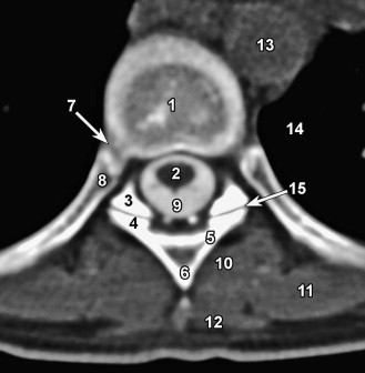
Thoracic Spine Anatomy | Radiology Key

Ct&Mri
Ct&Mri

Read Your MRI Basic Education from a World-Renowned Spine Expert |  ChiroGeek.com
Read Your MRI Basic Education from a World-Renowned Spine Expert | ChiroGeek.com

Axial CT scan demonstrating dosimetry for a lumbar spine lesion treated...  | Download Scientific Diagram
Axial CT scan demonstrating dosimetry for a lumbar spine lesion treated... | Download Scientific Diagram

Spine | SpringerLink

MRI Scans: Where Abnormal Findings Are Normal - Sohrab Gollogly MD

MRI Scans: Where Abnormal Findings Are Normal - Sohrab Gollogly MD

CT scan shows thecal sac compression without the destruction of bone or...  | Download Scientific Diagram
CT scan shows thecal sac compression without the destruction of bone or... | Download Scientific Diagram

Imaging of Spinal Metastatic Disease